Below is an example of what we are hoping to use.
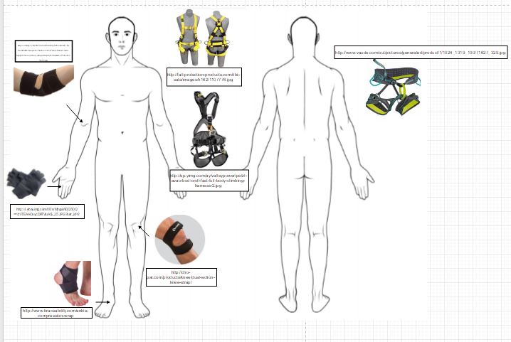 |
| This diagram shows the different braces we are looking into using, as well as the harnesses we are considering. If budget permits, we would like one the body harnesses shown, rather than the standard climbing harness on the right (which lacks the upper body section). |
 |
| Brainstorming on budgets and attachments. |
 |
| Isaac using Solidworks to create a CAD model for our project. |
"The influence of static and dynamic loading on marginal bone reactions around osseointegrated implants: an animal experimental study."
Link: http://onlinelibrary.wiley.com/doi/10.1034/j.1600-0501.2001.012003207.x/full
Although the bone-implant contact was not significantly different for the dynamically loaded versus the statically loaded and control implants, bone loss was observed at a close distance from the implants when they were loaded dynamically. This supports the hypothesis that excessive load can indeed trigger bone resorption through the induction of micro-damage in the bone.
"Mechanical Signaling for Bone Modeling and Remodeling"
Link: https://www.ncbi.nlm.nih.gov/pmc/articles/PMC3743123/pdf/nihms-281049.pdf
"Loading results in adaptive changes in bone that strengthen bone structure. Bone's adaptive response is regulated by mechanotransuction. We found that applying loading to the forelimbs of rats improved bone strength by 64%. Interestingly, the improvement in bone mineral content (BMC) in these rats was only a modest 7%. Consequently, mechanical loading adds mass but also causes the cross-sectional shape of long bones to become more structurally efficient." In adults (after growth has been cmpleted, "the effects of exercise shift from bone building to prevention of bone loss. In the adult skeleton, exercise reduces bone turnover by reducing bone resorption. Our studies employ a machine that applies axial loads to the forelimbs of rats or mice. In the rat ulna, where bone growth is directed to regions of high strain energy, improvement in function is dramatically improved. After 16 weeks of loading, the rat ulna will sustain 100-fold more loading cycles before failure." The article also includes information on the signaling pathways and specific cellular components/proteins that may cause bone growth. "Studying the effect of mechanical stimulation on resorption in animal models has been much more difficult than studying the effects on formation, and consequently, our understanding of the mechanisms involved is less developed. Part of the problem is that most of the animal models developed for studying the effects of increased loading on the skeleton are designed for addressing cortical bone in rodents. Because rodent cortical bone does not undergo Haversian remodeling, resorption data are scarce." It also says that bones may become desensitived to mechanical loading and produce less osteogenic activity. < This needs to be looked into more!!
"A threshold of mechanical strain intensity for the direct activation of osteoblast function exists in a murine maxilla loading model"
Link: https://goo.gl/S00H2u
"The purpose of this study is to clarify precisely what intensity level of mechanical strain is necessary to accelerate bone formation. The mice in the loaded group were subjected to continuous loading with 191kPa using the appropriate weight ingot for 30min/day on seven consecutive days." They tested for inflammation/swelling that resulted from the loading at two different locations: one close to the loading side and one farther away. They found that osteoclast number increased in both regions, but was much higher at the closer location.
"Bone Formation"
"As new bone material is added peripherally from the internal surface of the periosteum, there is a hollowing out of the internal region to form the bone marrow cavity. This destruction of bone tissue is due to osteoclasts, multinucleated cells that enter the bone through the blood vessels (Kahn and Simmons 1975; Manolagas and Jilka 1995). Osteoclasts are probably derived from the same precursors as macrophage blood cells, and they dissolve both the inorganic and the protein portions of the bone matrix (Ash et al. 1980; Blair et al. 1986). Each osteoclast extends numerous cellular processes into the matrix and pumps out hydrogen ions onto the surrounding material, thereby acidifying and solubilizing it‡ (Figure 14.17; Baron et al. 1985, 1986). The blood vessels also import the blood-forming cells that will reside in the marrow for the duration of the organism's life. The number and activity of osteoclasts must be tightly regulated. If there are too many active osteoclasts, too much bone will be dissolved, and osteoporosis will result. Conversely, if not enough osteoblasts are produced, the bones are not hollowed out for the marrow, and osteopetrosis results (Tondravi et al. 1997)."
"Benefits for Bone From Resistance Exercise and Nutrition in Long-Duration Spaceflight: Evidence From Biochemistry and Densitometry"
“These data document that resistance exercise, coupled with adequate energy intake (shown by maintenance of body mass determined by dual-energy X-ray absorptiometry [DXA]) and vitamin D, can maintain bone in most regions during 4- to 6-month missions in microgravity. This is the first evidence that improving nutrition and resistance exercise during spaceflight can attenuate the expected BMD deficits previously observed after prolonged missions. 2012 American Society for Bone and Mineral Research. It is well recognized by researchers in exercise physiology and bone biomechanics that bone needs to be optimally overloaded to have a stimulatory effect, which cannot always be provided by aerobic exercises."
"Rodent Research Contributes to Osteoporosis Treatments"
“We know that sclerostin production in bone is regulated by mechanical loading, but what we didn’t know was if you completely unloaded the skeleton’s bone formation pathway, which is essentially what happens in microgravity, whether our molecule that blocks sclerostin would still result in increased bone formation and bone strength. “If you take a human or an animal and put them in microgravity, they will lose muscle and bone at rates that are incredibly fast, much faster than a patient on Earth suffering from osteoporosis. They’ll lose bone at a rate 10 times faster in space than that individual. “For instance, static strains do not engender adaptive responses [8,9] whereas dynamic strains which change at high physiologic rates (as in impact loading) engender greater adaptive responses than those which change more slowly [10–13]. The on/off points therefore relate
to a strain-related stimulus rather than a particular strain value [9]."
Pharmaceuticals Solutions; "Mice Studies in Space Offer Clues on Bone Loss"
Link: http://www.nasa.gov/offices/oct/feature/mice-studies-in-space-offer-clues-on-bone-loss/
“One experiment focused on sclerostin, a naturally secreted protein that tells the body to dial down the formation of new bone. The mice were injected with an antibody that blocks sclerostin, essentially telling the body to “let up on the brake,” explains Chris Paszty, Amgen’s research lead on the project. That allowed the rodent bodies to keep regenerating bone tissue, resulting in increased mineral density and improved bone structure and strength. The results were encouraging: the mice injected with the antibody showed increased bone formation and improved bone structure and bone strength, similar to what was seen in the mice who remained on Earth.”
"Animal Experimental Measures of Functional Strain and Adaptation in Bone"
Source: Mechanical Strain and Bone Cell Function: A Review
P. J. Ehrlich and L. E. Lanyon
Department of Veterinary Basic Sciences, The Royal Veterinary College, London, UK
"These human exercise studies also support the data from animal studies that local loading induces local site specific changes in bone architecture [15–17]. Regardless of whether tensile or compressive forces were applied, the bone responded to intermittent, but not static loading. The first study to combine these approaches was that of O’Connor et al. [10] who used a pneumatic actuator to apply bending loads to sheep radii through metaphyseal pins. This study demonstrated a significant correlation between maximum strain rate and the degree of bone hypertrophy. This inference has been confirmed by a number of subsequent studies [11,19,32]."
No comments:
Post a Comment